
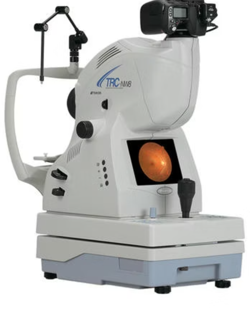
Fundus photography: a digital record of the retina and optic nerve
Retinal conditions can affect the macula, retinal blood vessels, vitreous or peripheral retina. Symptoms and urgency vary, so imaging must be linked with a dilated retinal examination.
What the test contributes
Fundus Camera provides objective information that can help the ophthalmologist document anatomy or function, compare findings over time and plan the next clinical step. The exact report fields and their significance vary with the instrument and the eye condition being evaluated.
What happens during evaluation
The visit usually begins with symptoms, medical history, vision assessment and eye examination. The doctor then decides whether this investigation is appropriate. Some tests are non-contact; others may require dilation, positioning, repeated measurements or additional examination. Patients should carry previous reports, current medicines and details of diabetes, blood pressure or earlier eye surgery.
How the report is interpreted
A result should not be interpreted in isolation. Image quality, test reliability, age, refractive power, corneal factors and previous treatment can influence the report. The specialist correlates the result with clinical findings and explains whether observation, repeat testing, medical treatment, laser, injection or surgery is being considered.
Questions patients can ask
- Why is this test being advised for my eye?
- What finding is the doctor looking for?
- Will the result change treatment or follow-up?
- Should this test be repeated, and after how long?
- Which warning symptoms require urgent review?
When urgent eye care may be needed
Sudden loss of vision, severe eye pain, chemical injury, eye trauma, new flashes with a curtain-like shadow, or rapidly increasing redness can require urgent in-person assessment. Online information cannot diagnose an emergency.
Meta Vision AI Lab pathway at SGVEH
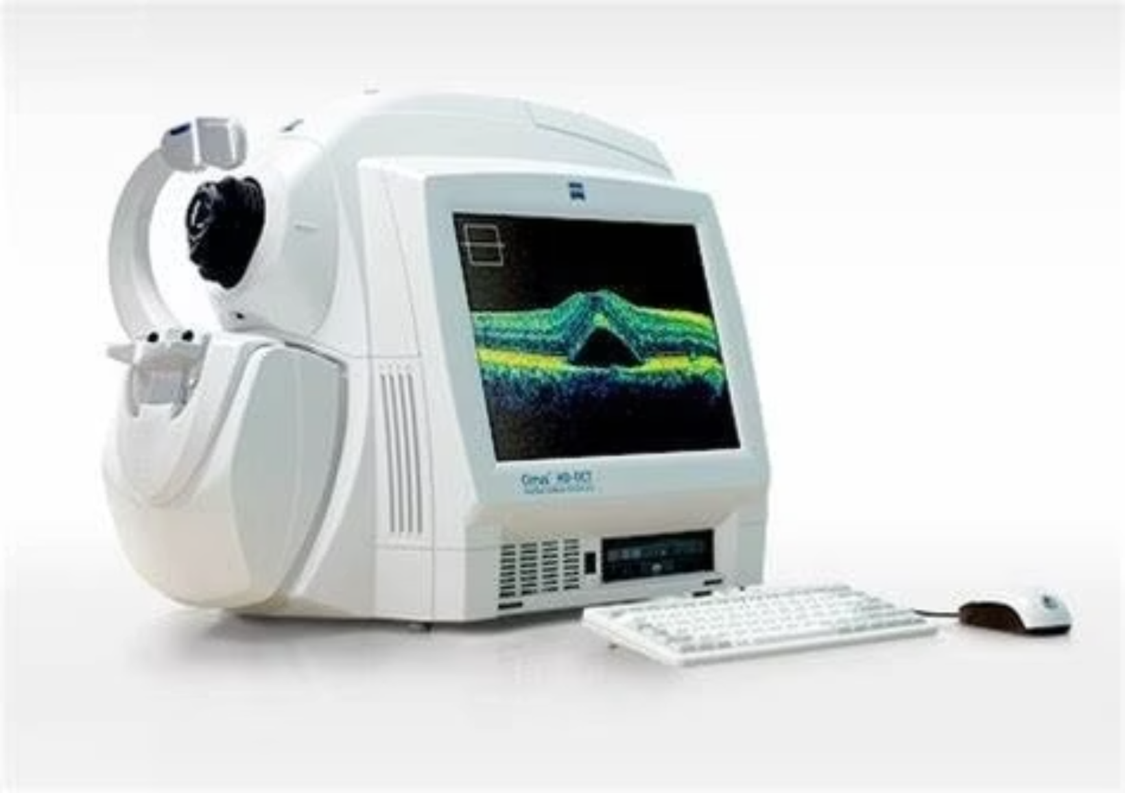
At Shri Ganesh Vinayak Eye Hospital, Raipur, advanced imaging is connected with the relevant retina, cornea, glaucoma, cataract, oculoplasty or comprehensive eye-care team. AI-assisted workflow can support analysis and organisation, while the final diagnosis and treatment decision remain with the ophthalmologist.
This article is general education and does not replace a personal eye examination.
This article is for general education and does not replace a personal eye examination. Diagnosis, suitability for treatment, risks and recovery vary by individual. Urgent symptoms require timely professional assessment.

 Team · Trust · Technology