
Corneal Transplant
Vision restoration for corneal scarring, opacity, and advanced corneal disease.
Shri Ganesh Vinayak Eye Hospital (SGVEH)
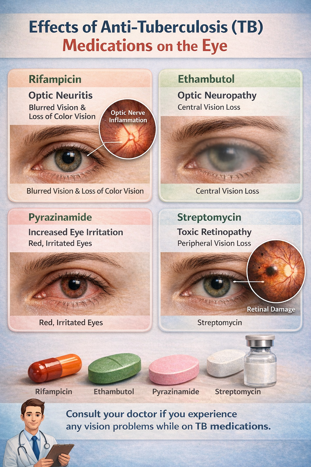
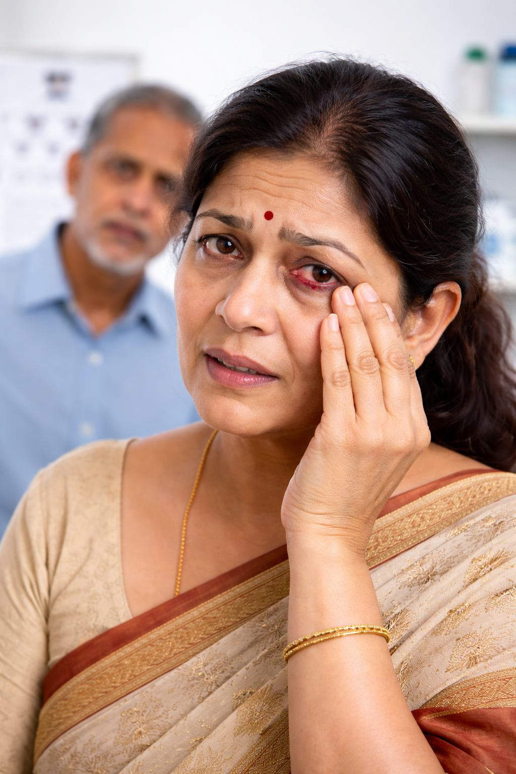
Patients undergoing TB treatment should have a baseline and periodic eye examination to ensure safe continuation of therapy.
Before starting TB medication.
Especially during Ethambutol therapy.
Detect early optic nerve toxicity.
Stop medication only after consulting physician.
Ethambutol is most commonly associated with optic nerve toxicity.
If detected early and medication is adjusted, vision may improve. Delay may cause permanent damage.
No. Immediately consult your treating physician and eye specialist before stopping medication.
At baseline and periodically during treatment, especially if taking Ethambutol.
Early screening prevents serious complications. Schedule an eye evaluation if you are on anti-TB therapy.
Shri Ganesh Vinayak Eye Hospital (SGVEH) • Comprehensive Eye Care
Patients with rheumatoid arthritis, lupus, ankylosing spondylitis, or other autoimmune conditions should undergo regular eye examinations to detect silent inflammation early.
Slit lamp evaluation and retinal assessment.
Anti-inflammatory drops or systemic treatment.
Artificial tears for chronic dry eye.
To prevent complications such as glaucoma or cataract.
Yes, it can cause dry eye, scleritis, and inflammation inside the eye.
Yes, untreated uveitis can lead to permanent vision loss.
Long-term steroid use may increase risk of glaucoma and cataract.
At least once a year, or more frequently if symptoms are present.
If you have arthritis or any autoimmune condition, do not ignore eye symptoms. Early evaluation can prevent serious complications.
Shri Ganesh Vinayak Eye Hospital (SGVEH) provides advanced cornea treatment and corneal transplant services in Raipur, including DSEK/DSAEK, DMEK, DALK and modern keratoconus management (C3R / corneal cross-linking). We support patients from across Chhattisgarh and Central India—including Korba, Champa, Jagdalpur, Dhamtari, Bilaspur—and nearby Odisha.

The cornea is the clear front layer of the eye. Cornea disease can cause blurred vision, pain, watering, redness, or whitening/scar. Many conditions are treatable—some need medicines, specialized lenses, or corneal transplant for vision restoration.
Sudden pain, redness, white spot, injury, or rapid vision drop can be urgent—please seek cornea evaluation quickly.
SGVEH cornea care focuses on accurate diagnosis, microscope-based evaluation, customized treatment plans and structured follow-up.

MBBS • DOMS • Cornea Consultant (LV Prasad Eye Institute)
Cornea • Corneal Transplant (DALK/DSAEK/DMEK) • Keratoconus (C3R)
Vision restoration for corneal scarring, opacity, and advanced corneal disease.
Partial-thickness transplant for endothelial problems (cornea swelling).
Advanced endothelial transplant technique for select candidates (high-quality vision recovery).
Lamellar transplant option for keratoconus/corneal scars where deeper layers are preserved.
Corneal cross-linking to strengthen cornea and slow progression in suitable cases.
Urgent diagnosis and treatment for microbial keratitis to prevent scarring and vision loss.
Chronic irritation, burning, watering—managed with step-wise therapy and follow-up.
Emergency care for scratches, metal foreign body, chemical injury and trauma.
Especially if there is light sensitivity, watering, or discharge.
May be corneal ulcer/infection—needs urgent evaluation.
Corneal swelling or scar can reduce clarity and contrast.
Can occur in keratoconus—early management protects vision.
We assess symptoms, prior surgery/injury/contact lens use, and test vision and eye pressure.
Detailed microscope exam checks corneal clarity, infection, scars and surface health.
For keratoconus and corneal shape evaluation, we may recommend corneal mapping and thickness tests.
Plan may include medicines, surface care, C3R, specialty lenses, or transplant depending on diagnosis.
For eligible cases, we counsel on the right transplant type (DSEK/DMEK/DALK) and ensure structured follow-up for healing and vision recovery.
A corneal transplant replaces diseased or scarred corneal tissue with healthy donor tissue to restore clarity and vision.
It depends on which cornea layer is affected. Endothelial problems may need DSEK/DMEK; keratoconus/scars may need DALK. A cornea specialist decides after examination.
Many keratoconus cases are managed with glasses, specialty lenses, and C3R (cross-linking) to slow progression. Some advanced cases may need transplant.
If you have severe pain, redness, light sensitivity, discharge, or a white spot, it may be an ulcer and needs urgent evaluation to prevent scarring.
Alert Shri Ganesh Vinayak Eye Hospital • Raipur
Just like high water pressure can burst a pipe, High Blood Pressure (Hypertension) damages the tiny, delicate blood vessels inside your eyes. This condition is called Hypertensive Retinopathy.

Early detection is the only cure.
Think of your eye's retina like a camera film. It needs a steady supply of blood.
When your Blood Pressure stays high (above 140/90) for a long time, the walls of the eye's blood vessels thicken to handle the pressure. Eventually, they become narrow or stiff.
The result? Blood cannot reach the retina properly, or the vessels burst (hemorrhage), causing vision loss.
Small blood vessels burst inside the eye. You might see red patches or dark floating spots (floaters) in your vision.
Just like a brain stroke, a clot can block an eye vein. This causes sudden, painless loss of vision in one eye. This is an emergency.
Fluid leaks into the 'Macula' (the center of your vision). Things will look blurry, wavy, or distorted (straight lines look bent).
High BP cuts off blood to the optic nerve (the cable connecting eye to brain). This can kill nerve cells and cause permanent blindness.
Note: In early stages, there are NO symptoms. That is why routine checkups are vital.
Dr. Charudutt Kalamkar uses advanced AIIMS-standard protocols to save your vision.
We take a high-resolution photo of your retina to see leaking vessels.
Like an MRI for the eye. It shows us swelling (fluid) inside the retinal layers.
If there is swelling or bleeding, we use Anti-VEGF injections or Green Laser to stop the leakage immediately.
Yes, possibly. BP damage starts at the outer edges of the retina where you don't notice it. By the time it affects your central vision (blurriness), the damage is already advanced. Diabetic and BP patients must get a checkup every 6 months.
No. Glasses only fix focus issues. High BP damages the "film" (retina) inside. If the film is damaged, changing the lens (glasses) won't help. You need medical retinal treatment.
If caught early (swelling/bleeding), we can restore much of the vision using injections. However, if the Optic Nerve strokes out (dies due to lack of blood), that vision cannot be recovered. Time is critical.
It is a sudden blockage of the retinal vein or artery. It usually happens to people with uncontrolled BP. If you suddenly lose vision in one eye, come to the hospital immediately (within 4 hours) for the best chance of recovery.
Shri Ganesh Vinayak Eye Hospital (SGVEH) • Raipur
Diabetic Retinopathy is a diabetes-related complication that damages the retina. High blood sugar levels cause retinal blood vessels to leak, swell, or close, leading to vision loss if not treated by a specialist.
At SGVEH Raipur, Dr. Charudutt Kalamkar (MS AIIMS, New Delhi) uses gold-standard technology to detect and treat diabetic eye diseases before they become permanent.
Diabetic retinopathy occurs when prolonged high blood sugar damages the microvasculature of the eye. It is the leading cause of vision loss in working-age adults. Initially, you may notice no changes, but as the condition progresses, it can cause bleeding inside the eye and retinal detachment.
The risk of developing retinopathy increases the longer you live with diabetes. Key factors include:
Risk increases significantly after 10–15 years of being diabetic.
Uncontrolled blood sugar (HbA1c > 7%) accelerates retinal damage.
High blood pressure adds strain to delicate retinal vessels.
Non-Proliferative (NPDR): Early stage where vessels leak fluid or fats into the retina.
Proliferative (PDR): Advanced stage where fragile new vessels grow and bleed into the vitreous.
Medicines like Lucentis or Avastin help reduce macular swelling and stop abnormal vessel growth.
Precision laser (Photocoagulation) seals leaking vessels and shrinks abnormal new growth.
A microsurgical procedure used for advanced cases involving vitreous hemorrhage or detachment.
Regular annual dilated eye exams are the most effective way to prevent 95% of diabetes-related blindness.
While advanced damage is permanent, early-stage NPDR can be managed and its progression halted through strict sugar control and specialist intervention.
No. We use numbing drops and very fine needles. Most patients describe it as a slight pressure sensation rather than pain.
Diabetic patients should have a comprehensive retinal exam at least once a year, or more frequently if any damage is detected.
Schedule your professional retina screening with Dr. Charudutt Kalamkar today at Shri Ganesh Vinayak Eye Hospital, Raipur.
Diabetes causes excess sugar to accumulate in your bloodstream. This damages the microscopic blood vessels in the Retina (the camera film of your eye). These vessels may leak fluid, causing swelling (Edema), or close entirely, starving the eye of oxygen.
Weakened blood vessels bleed into the eye. Leading cause of blindness in adults.
Fluid leakage in the center of vision (Macula) causes waviness and blurring.
High sugar levels cause the lens to cloud over faster than in non-diabetics.
Double the risk of high eye pressure, damaging the optic nerve silently.
Glaucoma (Kala Motia) is a progressive eye condition that damages the optic nerve, often due to high eye pressure. It is known as the "silent thief of sight" because it causes permanent vision loss without early symptoms.
Consult Dr. Charudutt Kalamkar (MS AIIMS, New Delhi)
Specialist in Glaucoma, Cataract & Neuro-Ophthalmology
In the early stages, Open-Angle Glaucoma has no warning signs. As the disease progresses, blind spots develop in your side (peripheral) vision. Regular checkups are the only way to catch it early.
Gradual loss of peripheral vision, leaving only central sight.
Seeing rainbow-colored circles around bright lights.
Sudden, severe eye pain accompanied by redness (Acute cases).
Upset stomach or vomiting linked to eye pressure spikes.
While vision loss cannot be recovered, we can prevent further damage using world-class technology.
Prescription eye drops to decrease eye fluid production or increase outflow.
Painless laser procedures (SLT/LPI) to open drainage angles and lower pressure.
Advanced surgical interventions like Trabeculectomy for complex cases.
No, glaucoma cannot be cured, and lost vision cannot be restored. However, it can be managed effectively to prevent blindness through early diagnosis and consistent treatment.
Most treatments, including lasers, are painless and performed in the OPD. Surgery is done under local anesthesia with minimal discomfort.
Glaucoma patients typically need checkups every 3 to 6 months to monitor eye pressure (IOP) and optic nerve health.
Painful swelling, redness, or lumps on your eyelid? Get expert diagnosis and relief. Trusted by patients across Raipur, Chhattisgarh & Odisha.
Direct WhatsApp: 7880177784
Eyelid infections occur when bacteria or blocked oil glands cause inflammation of the eyelid margin. In urban hubs like Raipur, Bilaspur, Durg, and Bhilai, factors like pollution and dust significantly increase cases of styes and chalazions.
Fig 1: Difference between a Stye (infection) and Chalazion (blockage).
Painful red bump caused by bacterial infection at the eyelash base.
Painless, firm lump due to a blocked oil gland further back on the lid.
Chronic inflammation causing crusty dandruff-like flakes on eyelashes.
Spreading infection causing entire eyelid swelling. Needs urgent care.
Common causes include:
*Warm compresses are the first line of defense.
Dr. Charudutt Kalamkar is a distinguished ophthalmologist trained at the prestigious AIIMS, New Delhi. With over two decades of experience, he leads the advanced eye care team at SGVEH.
Why Patients Trust Him: Known for his precise diagnosis and evidence-based approach to treating inflammatory eye conditions and infections.
Redness, discharge, or swelling in your child's eyes? Early diagnosis prevents long-term vision issues. Trusted by parents across Raipur, Chhattisgarh & Odisha.
Direct WhatsApp: 7880177784

Children are more vulnerable to eye infections because they frequently touch their eyes and play in close contact with others. In urban hubs like Raipur, Bilaspur, Durg, and Bhilai, factors like pollution, school outbreaks, and seasonal changes significantly increase cases of conjunctivitis and bacterial infections.
"Pink Eye" causing redness and sticky discharge.
Painful red bump on the eyelid due to blocked glands.
Itching and watering caused by dust or pollen.
Serious condition requiring immediate specialist care.
Most infections stem from:
*Prevention starts with hand washing.
Dr. Patel Bhavin is a renowned name in pediatric ophthalmology in Chhattisgarh. He specializes in treating delicate vision issues in infants and children with a gentle, child-friendly approach.
Why Parents Trust Him: He ensures accurate diagnosis avoiding unnecessary medication, focusing on the long-term visual development of your child.
Pediatric Care Shri Ganesh Vinayak Eye Hospital • Raipur
Spectacle Number (Refractive Error) in children happens when the eye cannot properly focus light on the retina. If left untreated, it can lead to lazy eye (Amblyopia), poor academic performance, and permanent vision issues.
80% of a child's learning is visual.
Think of the eye like a camera. The front of the eye (cornea and lens) focuses light onto the back of the eye (retina), which acts like the camera's film.
If the eyeball is slightly too long, too short, or the cornea is uneven, the light doesn't focus exactly on the "film." This makes the picture blurry. A spectacle lens simply bends the light before it enters the eye, fixing the focus.
Near-Sightedness: The child can see phones and books perfectly, but the blackboard or TV looks blurry. This is rapidly increasing due to screen time.
Far-Sightedness: The child struggles to focus on nearby objects like books or toys, often causing headaches or eye-crossing (squint).
Distorted Vision: The eye is shaped like a rugby ball instead of a football. It makes vision blurry or stretched at all distances.
Children rarely complain about bad vision because they think everyone sees the world exactly as they do. Watch for these clues:
Meet Our Pediatric Expert
When it comes to your child's vision, experience matters. Dr. Kalamkar specializes in diagnosing complex refractive errors and amblyopia (lazy eye) in children, ensuring they get the best start in life with clear vision right here in Raipur.
Staring at screens triggers the eyeball to grow longer (Myopia). We advise the 20-20-20 rule to reduce strain.
Outdoor play in natural sunlight releases dopamine, which stops the eye from growing too long. 1-2 hours daily is essential.
For rapidly increasing numbers, SGVEH offers Myopia Control Drops (Atropine) and special lenses to slow down progression.
No. While a healthy diet with Vitamin A is great for eye health, it cannot change the physical shape of the eyeball. Glasses are necessary to focus light correctly.
This is a myth. Glasses do not weaken the eyes; they help the brain see clearly. NOT wearing glasses can lead to "Lazy Eye" (Amblyopia).
By age 3 or 4, and definitely before starting school. We use special pediatric charts with shapes, so they don't need to know how to read.